The Human Brain
Glial Cells

| Functions of Glial Cells |
|---|
| Glial Cells occur in approximately the same numbers as neurones in most parts of the CNS. |
There are four main functions of glial cells:
|
| Astrocytes have processes that contact nerves cells, e.g. at synapses or nodes of Ranvier, and others that are in contact with blood vessels. This structure is thought to help regulate the supply of nutrients to the neurones and maintain local homeostasis of the environment of the neurones. |
| Oligodendrocytes provide the myelin wrapping of large axons in the CNS, and are therefore responsible for maintaining a high speed of conduction in central nervous pathways. |
| Microglia are the dormant phagocytes of the central nervous system, and can be activated to become phagocytes in these tissues |
| Ependymal cells are ciliated cells that line the ventricular system. |

| Astrocytes |
|---|
|
Astrocytes have numerous projections that anchor neurons to their blood supply. They appear to regulate the external environment of neurons by a selective movement of ions, e.g. potassium, and other chemicals.
Astrocytes are believed to be an essential part of the blood–brain barrier, and may release substances that regulate local blood flow, so that local blood flow is matched to the metabolic activity of neurones in that area. . |
| Astrocytes are able to take up various chemicals released at synapses and help to recycle them. Two examples would be the neurotransmitters glutamate or GABA, released during synaptic transmission, and recycled by conversion to glutamine, from which Glutamate and GABA are re-synthesised in the nerve terminal. |
| Astrocytes are believed to be an essential part of the blood–brain barrier, and may release substances that regulate local blood flow, so that local blood flow is matched to the metabolic activity of neurones in that area. |
| Astrocytes are connected to each other by gap junctions, and communicate using calcium and other chemical messengers |
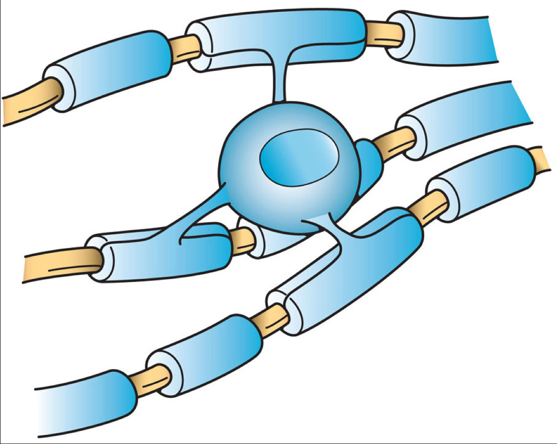
Oligodendrocytes are cells that coat axons in the CNS with many layers of their cell membrane that form the myelin sheath. The myelin sheath insulates the axon and is essential for fast transmission of nerve impulses.
The small distance between myelin sheaths is known as the node of Ranvier, and action potentials, the pulsatile short electrical signals that are conducted along the axon, jump from one node to the next, because of the high electrical resitance of multiple layers of cell membrane that consititute myelin.
Defects in this system occur in demyelinating diseases such as Multiple Sclerosis.
In contrast to the myelin of the peripheral nervous system, one oligodendrocyte can provide a myelin sheath for a number of axons.
Microglia: the dormant phagocytes of the CNS.
Microglia exist in several forms: they are normally present in an inactive form, but can be stimulated or primed to become active.
Active microglia are phagocytic and remove cell debris such as dead or damaged, degenerating neurones, bacteria and viruses.
The factors that trigger activation of Microglia include:
- viruses, bacteria and some CNS toxins.
- dead cells or cellular debris
- damaged neurones.
- other activated microglia.
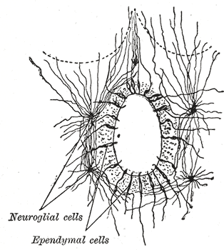
Ependymal cells line the cavities of the CNS and make up the walls of the ventricles. They have cilia that beat and help circulate the CSF and make up the Blood-CSF barrier.
Tanycytes are specialised ependymal cells found in the floor of the third ventricle; these cells have processes that extend deep into the hypothalamus and it is thought that they may convey signals from the CSF to the nuclei of the rostral ventral hypothalamus. They possibly have a role in the release of gonadotrophic hormone releasing hormone.